Bruker Light-Sheet Microscopes at Major Comprehensive Cancer Center
Bruker Corporation (Nasdaq: BRKR) has announced the installation of two MuVi™ and LCS SPIM™ light-sheet microscopes at Memorial Sloan Kettering Cancer Center (MSK). Funded by Cycle for Survival, these microscopes will enhance cancer research by allowing scientists to visualize cellular and tissue characteristics. The technology is expected to improve the understanding of cancer dynamics, facilitating the development of more effective treatment methods. This advancement underscores Bruker's commitment to supporting innovative cancer research.
- Installation of two light-sheet microscopes at Memorial Sloan Kettering Cancer Center enhances Bruker's product adoption.
- The funding from Cycle for Survival indicates strong community support for cancer research, potentially enhancing Bruker's reputation in the sector.
- None.
Insights
Analyzing...
Bruker Corporation (Nasdaq: BRKR), a leading supplier of single-plane illumination microscopy (SPIM) technology for research on live cells and cleared biological samples, today announced that two Luxendo MuVi™ and LCS SPIM™ light-sheet microscopes have been installed by Memorial Sloan Kettering Cancer Center (MSK). The funding for the two light-sheet fluorescence microscopes was supported by Cycle for Survival (https://www.cycleforsurvival.org). The new SPIM microscopes will help researchers visualize the cellular and tissue hallmarks of cancer and translate those findings into better cancer treatment methods.

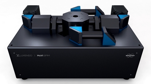
MuVi SPIM Multiview light-sheet microscope for live samples (Photo: Business Wire)
“By understanding how cells mobilize to build organs, researchers can glean insights into why some cells become cancerous and lead to organ destruction,” said Dr. Anna-Katerina Hadjantonakis, MSK Chair of the Developmental Biology Program. “Instruments such as these are useful for imaging across differing length scales — from subcellular to single cells to tissue-level processes — allowing researchers to study cellular dynamics and cellular motion, processes that enable cells to metastasize.”
“Light-sheet fluorescence microscopy has emerged as a uniquely powerful method for high-resolution, cleared-sample and dynamic biological imaging,” added Dr. Lars Hufna
FAQ
What new technology has Bruker Corporation installed at Memorial Sloan Kettering Cancer Center?
How is the funding for Bruker's new microscopes sourced?
What is the significance of the new microscopes for cancer research?
What company is associated with the stock symbol 'BRKR'?


